Art-labeling activity structure of a skeletal muscle fiber unveils the intricate world of muscle biology, providing a comprehensive framework for visualizing and analyzing the building blocks of movement. This detailed guide delves into the histological features of muscle fibers, the principles of art-labeling techniques, and the strategies for labeling specific muscle fiber types.
By exploring the applications of art-labeling in skeletal muscle research, we gain a deeper understanding of muscle development, regeneration, and disease.
Histological Features of Skeletal Muscle Fiber

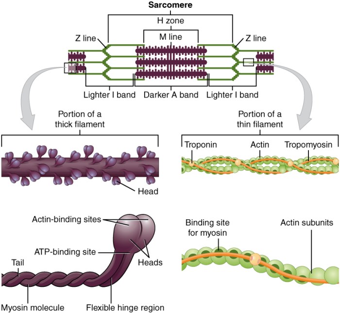
Skeletal muscle fibers are elongated, cylindrical cells that are responsible for voluntary movement. They are composed of multiple nuclei located beneath the sarcolemma, the cell membrane of muscle fibers. The cytoplasm of skeletal muscle fibers is filled with myofibrils, which are long, cylindrical structures that contain the contractile proteins actin and myosin.
Myofibrils are organized into repeating units called sarcomeres, which are the basic units of muscle contraction. Each sarcomere is composed of a thick filament (myosin) and two thin filaments (actin).
Art-Labeling Techniques for Skeletal Muscle Fibers

Art-labeling techniques are used to visualize specific components of skeletal muscle fibers. These techniques involve the use of antibodies or fluorescent probes that bind to specific proteins or structures within the muscle fiber. Immunohistochemistry is a commonly used art-labeling technique that involves the use of antibodies to detect the presence of specific proteins.
Fluorescent labeling involves the use of fluorescent probes that emit light when they bind to specific molecules.
Labeling Strategies for Specific Muscle Fiber Types

Different types of skeletal muscle fibers can be labeled using specific antibodies or fluorescent probes. Type I fibers, which are slow-twitch and fatigue-resistant, can be labeled using antibodies against slow myosin heavy chain. Type IIa fibers, which are fast-twitch and fatigue-resistant, can be labeled using antibodies against fast myosin heavy chain type IIa.
Type IIx fibers, which are fast-twitch and fatigable, can be labeled using antibodies against fast myosin heavy chain type IIx.
Analysis of Art-Labeled Skeletal Muscle Fibers: Art-labeling Activity Structure Of A Skeletal Muscle Fiber
Art-labeled skeletal muscle fibers can be analyzed using a variety of techniques, including image acquisition and processing. Image acquisition involves capturing images of the labeled muscle fibers using a microscope or other imaging device. Image processing involves enhancing and analyzing the images to quantify muscle fiber size, shape, and distribution.
Applications of Art-Labeling in Skeletal Muscle Research
Art-labeling techniques have a wide range of applications in skeletal muscle research. These techniques can be used to study muscle development, regeneration, and disease. For example, art-labeling has been used to identify the different types of muscle fibers that are present in different muscles and to study how these fibers change in response to exercise or injury.
FAQ Summary
What are the key histological features of a skeletal muscle fiber?
Skeletal muscle fibers are characterized by their striated appearance, the presence of myofibrils, and the arrangement of nuclei at the periphery of the fiber.
How does art-labeling help visualize skeletal muscle fibers?
Art-labeling techniques, such as immunohistochemistry and fluorescent labeling, use specific antibodies or fluorescent probes to bind to target proteins within muscle fibers, allowing for their visualization and analysis.
What are the different strategies for labeling specific muscle fiber types?
Specific muscle fiber types can be labeled using antibodies or fluorescent probes that target unique proteins expressed in those fiber types, such as type I, type IIa, and type IIx fibers.